
Updated: 2024-04-30
On April 23, Associate Professor Xiao Zhenyu, of the School of Life Science at Beijing Institute of Technology (BIT), alongside researchers Wang Hongmei, Yu Leqian, and Guo Jingtao from the Institute of Zoology (IOZ) at the Chinese Academy of Sciences (CAS), Professor Wei Yulei from China Agricultural University, and Assistant Researcher He Nannan from the First Affiliated Hospital of Zhengzhou University, published an article in the renowned international academic journal Cell, titled "3D Reconstruction of a Gastrulating Human Embryo." Using chip technology and 3D animation reconstruction, they reconstructed a high-resolution 3D digital human gastrula for the first time, filling in the knowledge gap regarding human embryonic development during the gastrula stage. This research provides important information for understanding human early embryonic development and related diseases, laying a scientific foundation for improving adverse pregnancy outcomes.

Abnormalities during gastrulation are associated with various pregnancy and development-related diseases, such as recurrent pregnancy failure, congenital heart disease and neural tube defects. Several research teams have analyzed the events of human embryonic development from 0 to 14 days after fertilization, with the gastrula stage encompassing 14 to 21 days after fertilization. During this stage, the formation of the three germ layers gives rise to all the organs and systems in the human body. However, due to the scarcity of samples, critical knowledge about the cell lineages, signaling pathways, precise spatial locations and the role of extraembryonic tissues in gastrulation has remained elusive.
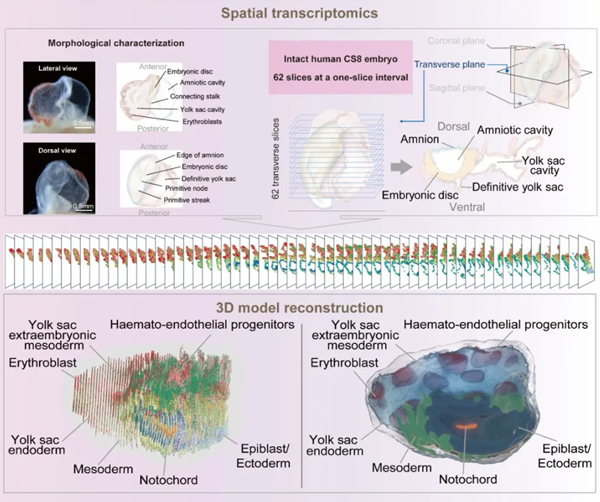
Flowchart for 3D reconstruction of human gastrula [Photo/english.bit.edu.cn]
Primates, such as monkeys, share significant physiological and embryological similarities with humans, providing an excellent model for studying key events and mechanisms during gastrulation. Through the joint efforts of teams led by Chinese scientists Ji Weizhi, Wang Hongmei and Peng Guangdun, alongside other international research teams, significant scientific questions about the gastrula stage in monkeys are being systematically resolved. However, to truly understand the formation of the human gastrula, direct studies on human embryos are essential. This study systematically depicts the embryonic morphology, cell clusters, spatial locations and transcriptional features, elucidating the development of key extraembryonic tissues, including amniotic cells and the yolk sac hematopoietic lineage.
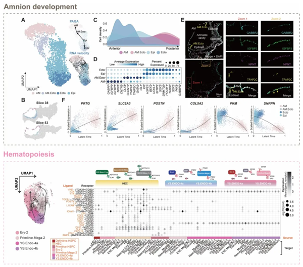
Development of the amnion and hematopoietic specialization of the yolk sac in the human gastrula [Photo/english.bit.edu.cn]
Additionally, the study analyzes key signaling pathways along the anterior-posterior, dorsal-ventral and left-right axes during gastrulation, proposing the possibility of a new signaling source and an "organizer" in the embryo's tail region. This offers an important glimpse into early human embryonic development.
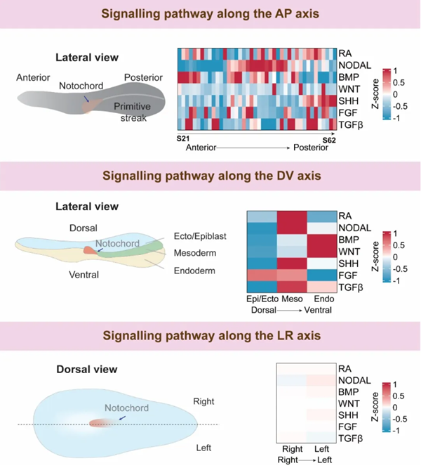
Key signaling pathways for anterior-posterior, dorsal-ventral, and left-right body axis in the human gastrula [Photo/english.bit.edu.cn]
About the Author
Xiao Zhenyu is an associate professor, a doctoral advisor and a team member with Professor Dong Lei at the School of Life Science at BIT. He has published 17 SCI papers in renowned international academic journals, including Cell, Nature Genetics, Developmental Cell, Nature Communications, and Protein Cell, with a total impact factor of 243 and 449 total citations. He has led projects funded by the National Natural Science Foundation of China (Youth Project and General Program), Horizon Europe's Marie Skłodowska-Curie Actions (MSCA), and participated as a key member in the Class A strategic pilot technology projects of the Chinese Academy of Sciences. Xiao has also received Horizon Europe MSCA’s outstanding talent award and the first prize in the Science and Technology Awards for Maternal and Child Health for his research achievements.


